Medical applications - a cancer patient has a new femur built using 3D prints
This real-life story begins with a young man who had a malignant tumour in his femur. The removal and repair of the tumour required a very challenging surgical procedure. The doctors decided to repair the femoral defect using the patient's own vascularized fibula, which is worked into the donor femoral component. The surgical method is known as the Capanna technique. The patient's treatment was planned and carried out at Tampere University Hospital.
The specialist in charge of planning and implementing the patient's treatment explains:
- Before the surgery, we prepared a fibula graft to be implanted in the patient's femur. This structure was supported by a long plate that was attached to the outer edge of the thigh. The great thing about this solution was that it saved the patient's own knee and hip joints.
The operation required great precision and careful planning. A 3D printed model of the femur was used, which helped the surgeon to accurately plan the position of the plate and its correct alignment with the patient's femur before the actual surgery. The accurate measurement of the fibula also helped to outline how the patient's own femoral heads and donor bone should be treated for the best outcome.
The 3D prints were made using HP's Jet Fusion printing technology, based on data generated by computed tomography. Another important part of the collaboration was to ensure that confidential patient data was transferred securely between the hospital and the 3DStep service.
- Although 3D prints are obviously not the same as live bones, they provided a valuable opportunity to dry run the surgery in advance," says the specialist.
The key to success was the close collaboration between the surgeon and the technician to ensure that the prints exactly matched the needs of the surgery. For surgeons, the importance of collaboration is particularly important when it comes to using 3D-printed, patient-specific sawing guides.
Overall, 3D prints can be an invaluable tool for planning and performing complex surgeries. For example, in the case described above, the surgeons were able to go through the steps of the surgery together with the physical models.
- The operation was very challenging at all stages and the 3D prints made it much easier to visualise the different phases. 3D prints made it particularly easy to plan and carry out the reconstruction of the femur. The models were produced quickly and were of excellent quality.

After tumour removal, a 17 cm gap in the middle of the femur was left, which had to be replaced with a load-bearing structure. There is no good prosthetic reconstruction available for the tubular shaft, so the aim was to achieve as biological a reconstruction as possible. The replacement used the patient's own fibula, which was machined into the femur. The reconstruction was further supported by a long plate on the outer edge of the femur.
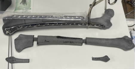
The femoral model allowed the patient's plate to be bent into a fully articulated position a week before the operation. This provided one additional reference for the femoral position during the procedure: if the plate is well positioned, the femoral axis, rotation and dimension should be approximately correct. In addition, an accurate calibre of the fibula was obtained, which provided a guideline for how the patient's own femur and the donor femur should be treated in order to implant the fibula into the ends of the severed femur.

The 3D prints were produced using HP's Jet Fusion printing technology from dedicated computed tomography scans. The prints were made of the femur in two pieces and one of the fibula. The data transfer to 3DStep was carefully planned and implemented with the help of an IT specialist.